New ‘molecular magnifying glass’ illuminates critical points in proteins to track Alzheimer’s disease
A team at Rice University has developed a strategy to magnify small segments of proteins within living cells.
The method reveals subtle environmental changes that may signal the early stages of diseases such as Alzheimer’s, Parkinson’s, and cancer. The findings also point to new ways of screening drugs that target protein aggregation disorders.
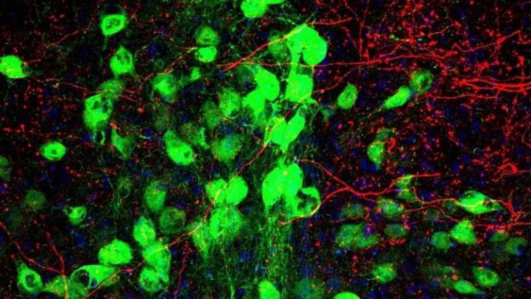
The researchers created a fluorescent probe, called AnapTh, in precise protein subdomains. This probe monitors real-time changes that conventional techniques often fail to detect.
By tracking localized changes, scientists can observe how distinct regions of the same protein behave differently as they begin to aggregate.
“Essentially, we built a molecular magnifying glass,” said Han Xiao, professor of chemistry and director of Rice’s SynthX Center. “This allows us to visualize subtle environmental changes that previously went unnoticed.”
The team based their design on the idea that initial local changes appear before visible agglomeration.
AnapTh, a fluorescent amino acid, changes its emission spectrum depending on the surrounding microenvironment. Using genetic code expansion, the scientists inserted the probe into chosen locations without disrupting the folding or function of the protein.
This approach provided spatial resolution and real-time monitoring that existing tools could not offer.
“We wanted a method to illuminate just one spot on a protein and observe what happens around it in living cells,” said Mengxi Zhang, a graduate student and co-lead author. “When aggregation begins, some parts become denser and more hydrophobic, while others remain unchanged.”
Applying the technique to disease-related proteins revealed a surprising pattern. Aggregation did not occur uniformly.
Some subdomains showed increased fluorescence intensity and spectral changes, indicating denser and chemically altered environments. Others remained unchanged.

This discovery challenges older models that described aggregation as uniform.
Instead, evidence shows that aggregation begins at discrete “hot spots” and then spreads. These initial localized unfolding events could become future biomarkers or serve as therapeutic entry points.
The results reshape how scientists view protein aggregation.
They highlight a process that is uneven and dynamic, with specific locations driving the first disease-related changes. This new perspective provides a fresh insight into the molecular triggers of neurodegeneration.
Protein aggregation underlies devastating diseases such as Alzheimer’s plaques, Parkinson’s Lewy bodies, and misfolded proteins in cancer.
Understanding where and how these changes begin gives researchers a clearer path to treatments. The discovery offers a sharper lens for studying how small errors in folding a helix turn into major health threats.
The Rice team also demonstrated how the platform could aid in drug discovery. By detecting early changes in the subdomain, the tool can track disease progression with greater sensitivity.
Researchers can also identify compounds that intervene before aggregation spreads.
“This platform gives us a head start,” said Shudan Yang, a graduate student and co-lead author. “Now we can test potential inhibitors and see, at the first sign of trouble, if they prevent the incorrect local unfolding.”
That level of precision, Yang added, is exactly what drug development needs.
The approach could shorten drug screening times and improve targeting of specific disease weaknesses.
Co-authors of the study include Rice researchers Shikai Jin, Yuda Chen, Yiming Guo, Yu Hu, and Peter Wolynes.
The study was published in the journal Nature Chemical Biology.