Mapping the intricate universe of the human brain
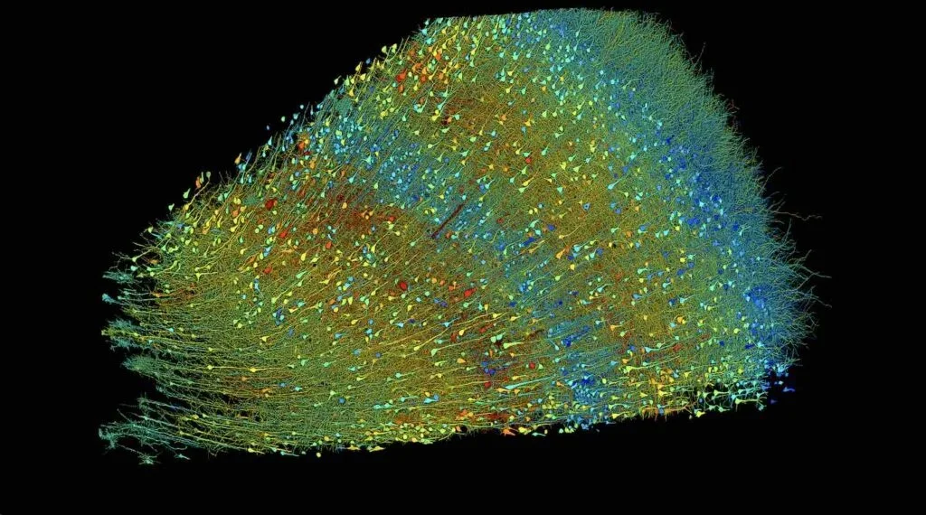
In a scientific achievement, researchers at Harvard University and Google have produced the most detailed map ever created of the human brain. Their collaborative effort, over a decade in the making, has revealed the staggering complexities of neural connections and circuitry within just a tiny cubic millimeter sample of brain tissue.
The study, led by Harvard’s Jeremy R. Knowles Professor of Molecular and Cellular Biology Jeff Lichtman, leveraged cutting-edge electron microscopy imaging techniques coupled with powerful artificial intelligence from Google. The result is an unprecedented, high-resolution 3D reconstruction offering novel insights into the intricate inner workings of the human brain.
Within that single cubic millimeter of temporal cortex tissue, taken from the brain of an epilepsy patient, lie almost 57,000 cells along with hundreds of thousands of synaptic connections between neurons. Mapping these minuscule structures in such fine detail produced a staggering 1,400 terabytes of data – a scale highlighting both the incredible complexity of our brains and the prowess of modern imaging and AI capabilities.
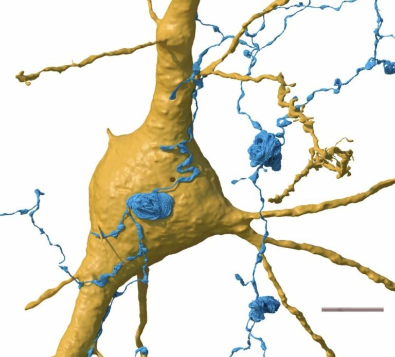
As Lichtman describes it, the visualization reveals “an alien world inside your own head.” The paper’s authors point out that while we recognize the brain’s neural circuits as representing the crucial distinction between humans and other lifeforms, our understanding of this synaptic circuitry has remained fairly opaque until now.
To create the map, termed a “connectome” by the researchers, Lichtman obtained a well-preserved tiny brain sample over 10 years ago during an epilepsy surgery. Using one of the most powerful electron microscopes, they imaged the sample down to a resolution of just 8 x 8 x 30 nanometers per pixel.
From this raw image data, Google’s Connectomics team applied advanced machine learning algorithms to computationally reconstruct and distinguish individual neurons, synapses, blood vessels, and other brain structures throughout the tissue sample. The visualizations revealed a whole new class of neurons hidden in the deep layers of the brain, as well as “very powerful and rare” connections between neurons that extend over long distances. This data has been made available online, along with tools to help other researchers analyze it.
“This work provides evidence of the feasibility of human connectomic approaches to visualize and ultimately gain insight into the physical underpinnings of normal and disordered human brain function,” wrote the researchers in their newly published Science paper. To enable further study, they have made the full dataset and analysis tools freely available online.
Previous attempts to map the human brain at this scale faced limitations, as brain biopsies rarely occur outside of tumor removal, and lab-grown organoids lack the full cellular organization of real brain tissue. The new study circumvented these issues by using surgically-removed yet still healthy tissue.
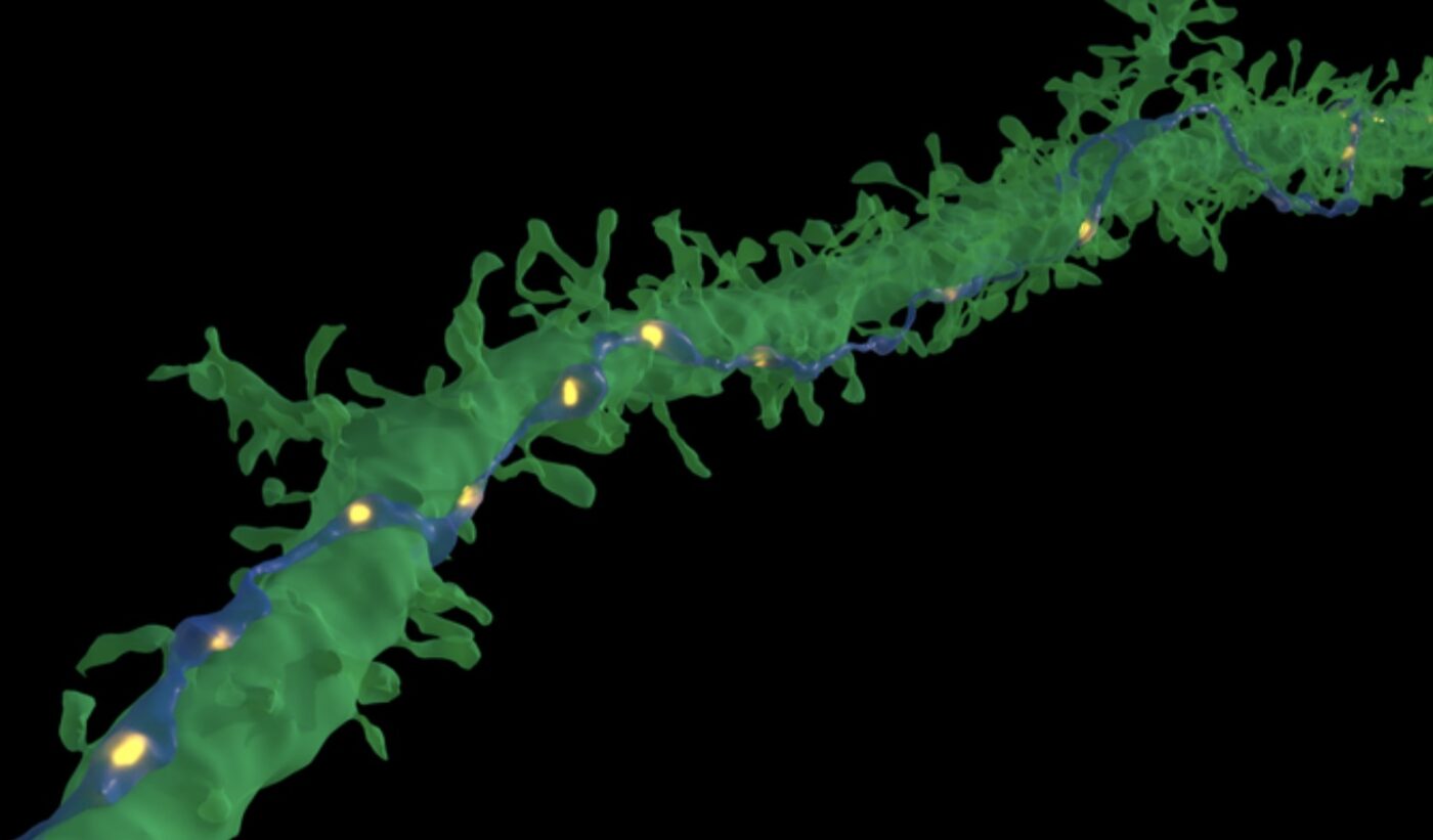
The endeavor is part of a broader effort funded by the NIH BRAIN Initiative, with an ultimate goal of producing a complete neural wiring diagram or “connectome” of an entire mouse brain at nanoscale resolution. Achieving this will require imaging and reconstructing approximately 1,000 times more data than the 1 cubic millimeter sample mapped in the current study.
While an entire human brain at such fine resolution may be impractical with current techniques, the new visualizations have opened up a window into the neurological “alien” universe within our heads. The public dataset provides an invaluable resource that could enable myriad new discoveries about how our neural circuitry gives rise to human cognition, perception, and brain disorders.
“Given the enormous investment put into this project, it was important to present the results in a way that anybody else can now go and benefit from them,” said Google Research collaborator Viren Jain, speaking to the transformative potential of freely sharing such pioneering work.
- See also: New method of producing human cartilage
After centuries of studying the brain mainly through static histological sections, neuroscientists can now virtually inhabit and explore entire neural landscapes in unprecedented detail. This hidden neural world has become visible, and a new frontier of brain research is approaching.

