Mucus-based bioink for printing and growing lung tissue
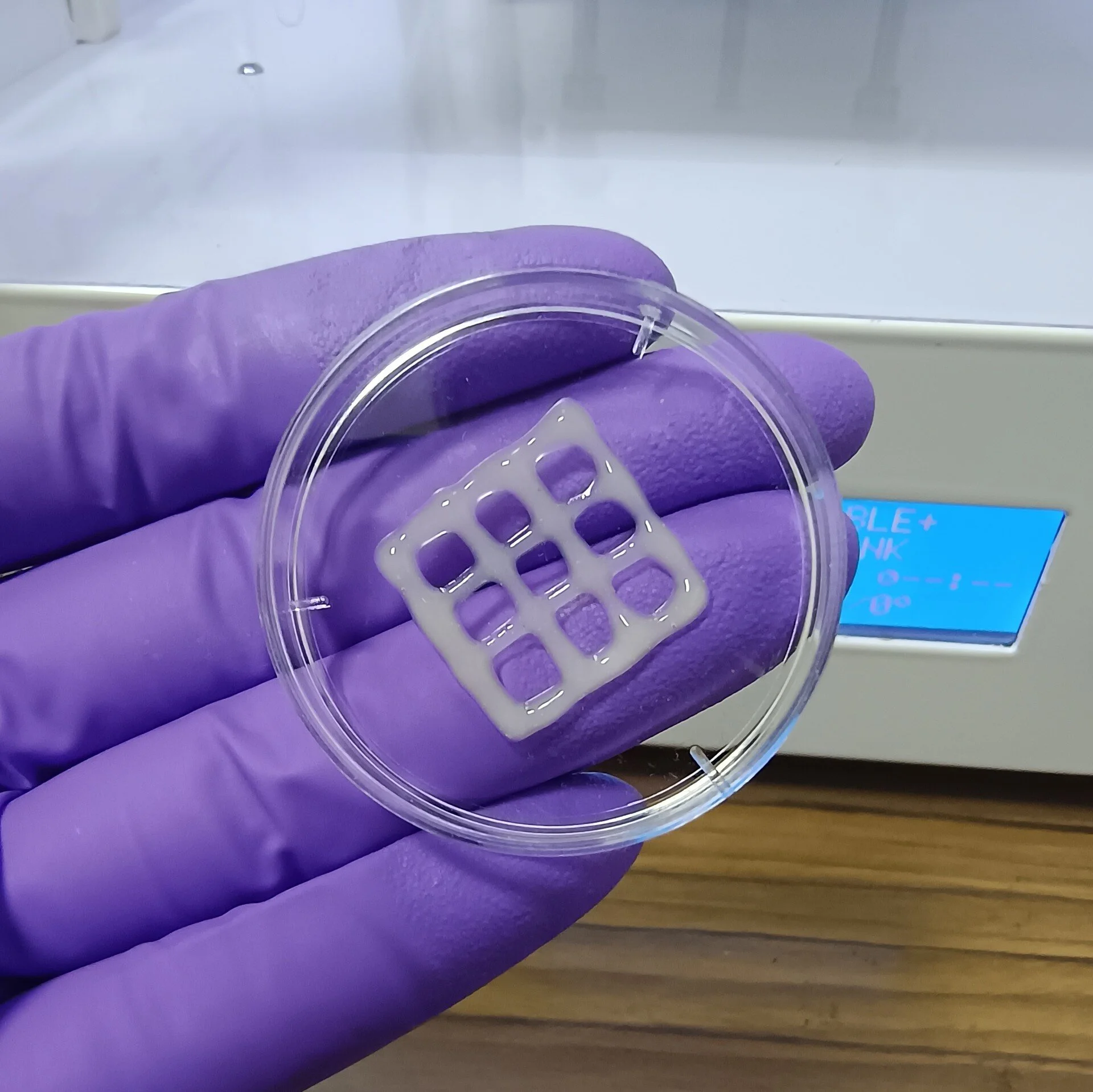
It’s so sad to think that lung diseases are responsible for millions of deaths worldwide every year. Despite the amazing advances in medicine that we’ve seen, treatment options remain limited and animal models for studying these diseases and testing drugs are often inadequate. But there’s some really exciting new research published in the journal ACS Applied Bio Materials that could represent a significant advance. Scientists have developed a mucus-based bioink for 3D printing lung tissue, which is an amazing innovation that could transform the study and treatment of chronic lung diseases.
Current Limitations and the Need for New Models
For individuals afflicted with severe pulmonary disorders, organ transplantation represents a potential avenue for treatment. However, the scarcity of available donors constrains the feasibility of this approach. In the meantime, pharmacological and other therapeutic interventions are capable of controlling the symptoms associated with these conditions, but they do not constitute a cure for diseases such as chronic obstructive pulmonary disease (COPD) and cystic fibrosis. Researchers frequently utilize rodent models to develop novel pharmaceuticals. However, these models often fail to fully capture the intricacies of human lung diseases, thereby impeding the accurate prediction of new treatments’ safety and efficacy.
3D Bioprinting: A New Frontier
The field of bioengineering has been engaged in the pursuit of developing a more accurate alternative to animal models through the creation of lung tissue in laboratory settings. Three-dimensional printing of structures that mimic human tissue represents a promising technique; however, the development of a suitable bioink that supports cell growth represents a significant challenge. Ashok Raichur and his team have addressed this challenge in an innovative manner.
The Mucus-Based Bioink
The Raichur team made an exciting discovery when they used mucin, a component of mucus, for bioprinting. This was a new and promising avenue that hadn’t been widely explored before. Mucin has segments of molecular structure that resemble epidermal growth factor, a protein that helps cells stick together and grow. To create the biotin, the researchers had a brilliant idea! They reacted the mucin with methacrylic anhydride to form methacrylated mucin (MuMA), which they then mixed with lung cells.
To make the biotint nice and thick and help the cells stick and grow, the researchers added hyaluronic acid, which is found in our bodies. After printing the ink in test patterns, such as round and square grids, the structure was exposed to blue light to cross-link the MuMA molecules, which made it into a porous gel that can soak up water and support cell survival.
Promising Results
The tests yielded some amazing results! It was found that the interconnected pores in the gel played a crucial role in facilitating the diffusion of nutrients and oxygen, which are essential for cell growth and the formation of lung tissue. The printed structures were non-toxic and biodegraded slowly under physiological conditions, making them perfect for implants where the printed support would gradually be replaced by newly grown lung tissue.
This amazing biotint could also be used to create 3D models of lungs, which would be invaluable for studying lung diseases and evaluating new treatments.
This is a truly groundbreaking moment in the field of bioengineering! The creation of a mucus-based bioink for 3D printing lung tissue is a game-changer. It offers a new and precise way to study lung diseases and opens the door to future treatments that could improve the quality of life of millions of people affected by these debilitating conditions. With further research and development, 3D printing of human tissue could become a crucial tool in regenerative medicine and the treatment of chronic diseases.
The research was published in ACS Applied Bio Materials.

