20 jaw-dropping images of the microscopic world around us
Byomniletters.com
Explore the top 20 award-winning images from the 50th Nikon Small World Photomicrography Competition.
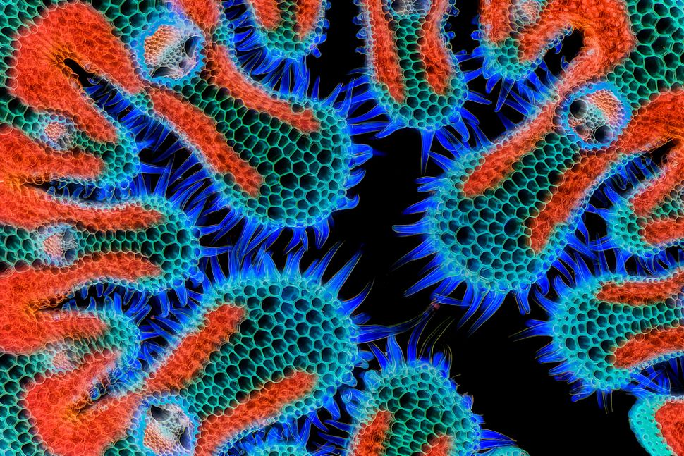
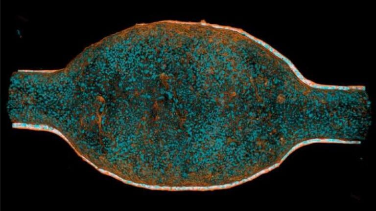
A microscopic image of a cannabis plant won third place at the Nikon Small World Photomicrography Competition. (Image credit: Chris Romaine/Nikon Small World 2024)
A panel of judges selected 20 winning entries, chosen from approximately 2,100 submissions, for their exceptional clarity in capturing the minutest details of their subjects.
Eric Flem, senior manager of CRM and communications at Nikon Instruments, remarked in a statement shared with Live Science, “Sometimes, we overlook the tiny details of the world around us. Nikon Small World serves as a reminder to pause, appreciate the power and beauty of the little things, and to cultivate a deeper curiosity to explore and question.”
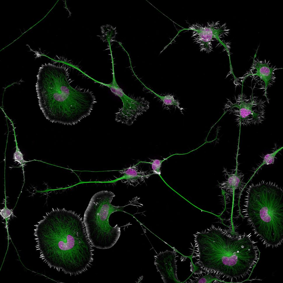
Bruno Cisterna and Eric Vitriol, researchers from the Department of Neuroscience and Regenerative Medicine at Augusta University in Georgia, earned first place with their image of differentiated mouse brain tumor cells. This image highlights how alterations in the cell’s cytoskeleton, which maintains cell structure and function, can contribute to neurodegenerative disorders such as Alzheimer’s disease.
Advertisement - Continue Reading Below
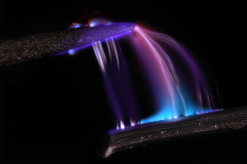
Astronomer-turned-photographer Marcel Clemens received second place for his image of an electrical arc between a pin and wire, while cannabis photographer Chris Romaine secured third place for his photograph of a cannabis plant leaf.
The full gallery of the 20 winning images can be viewed below.
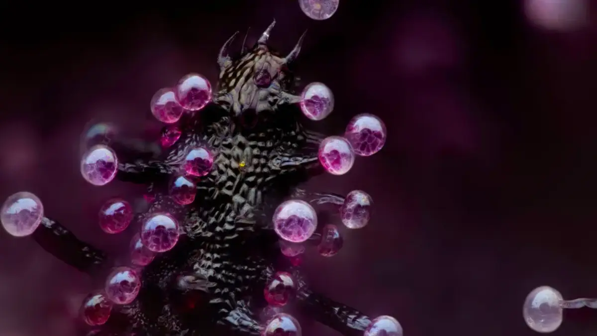
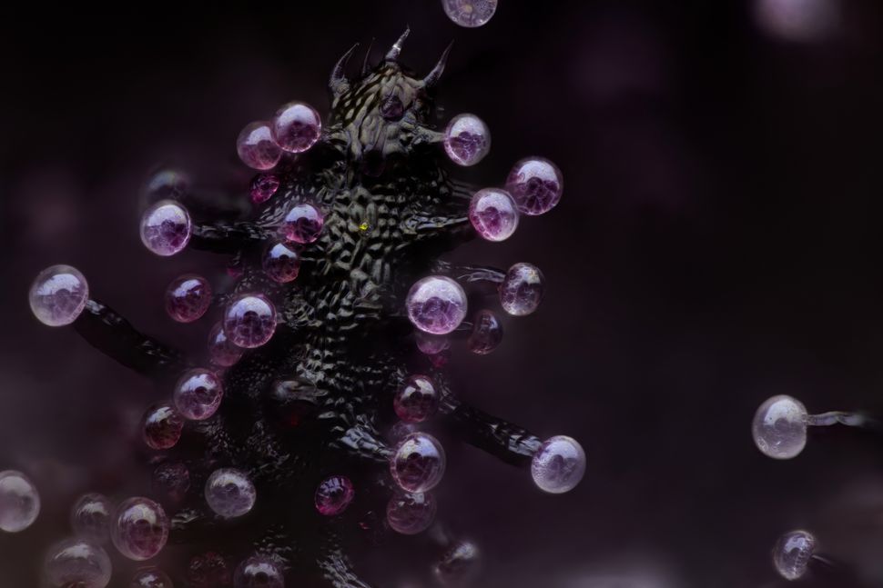
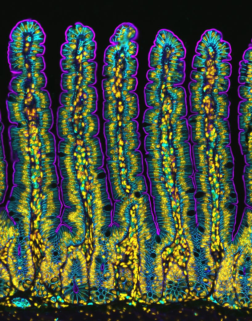
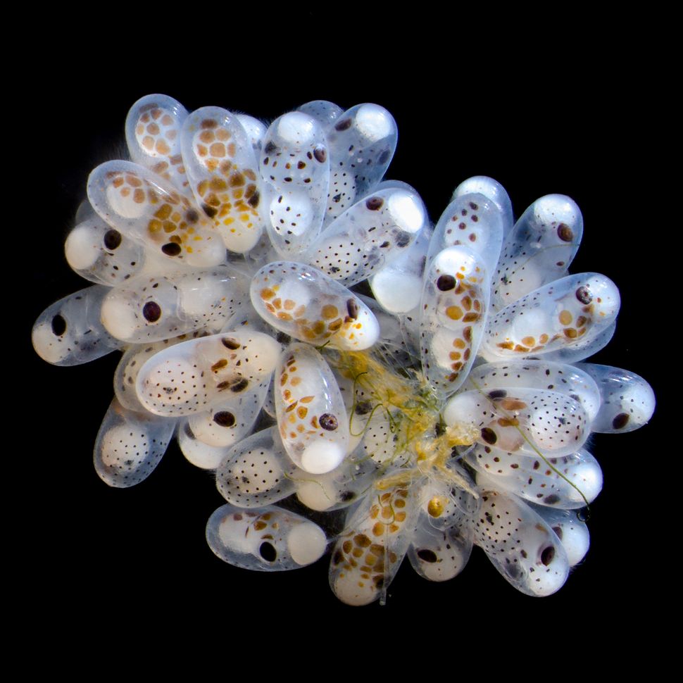
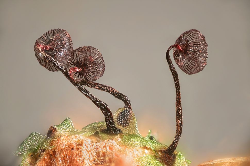
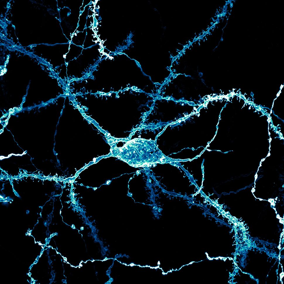
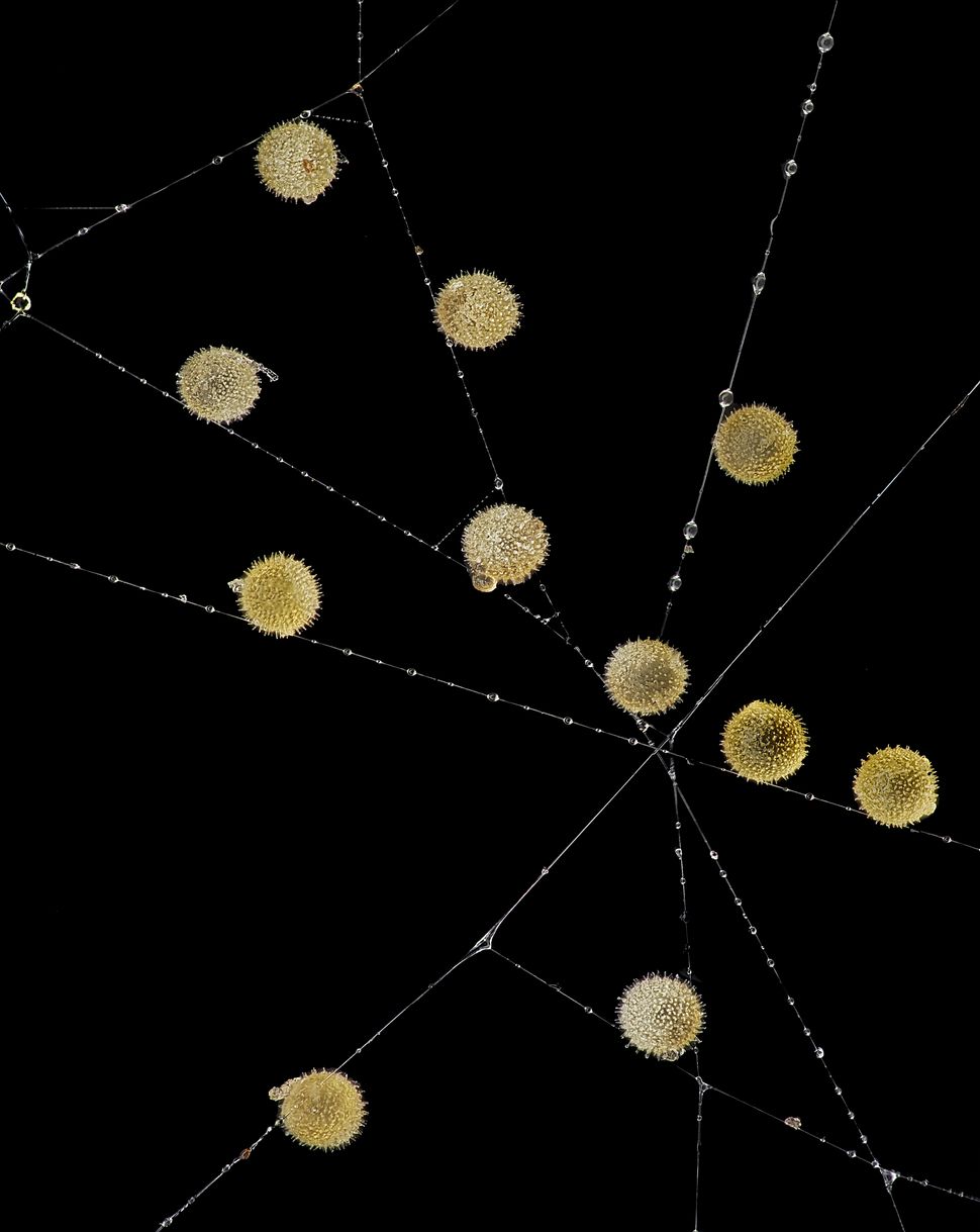
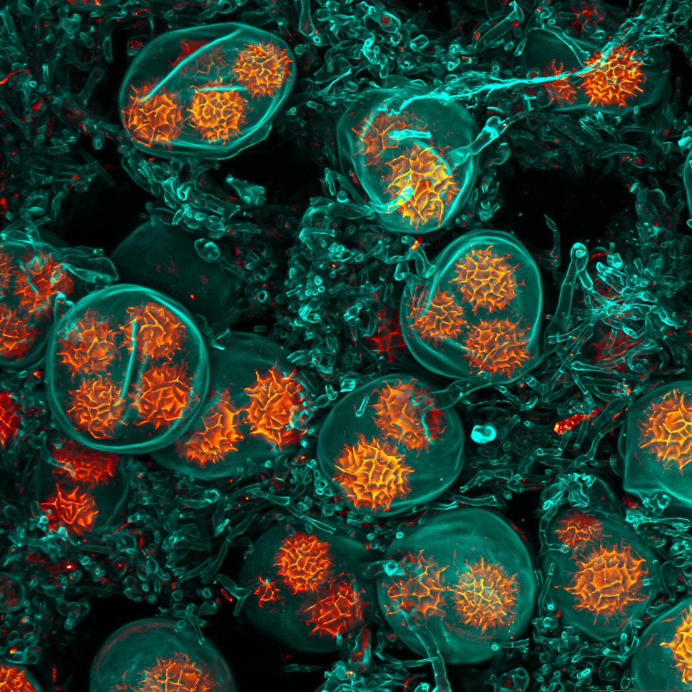
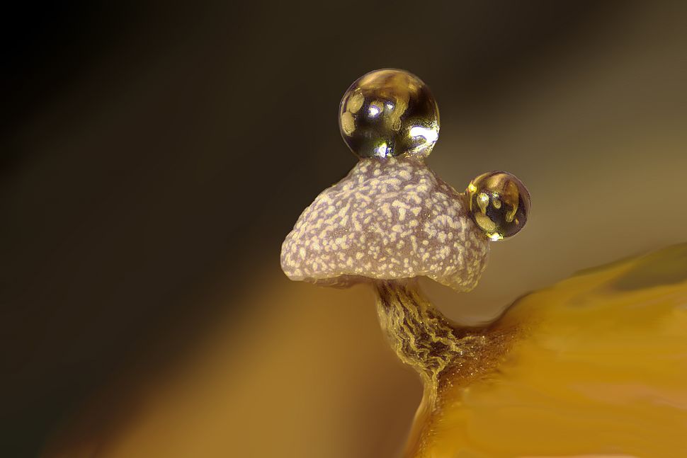
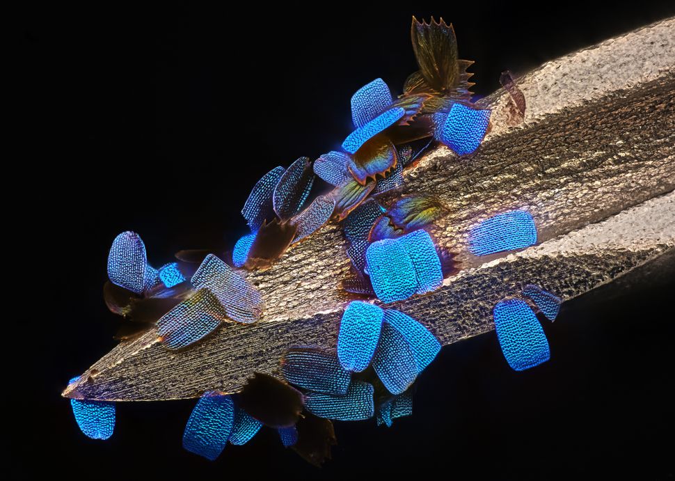
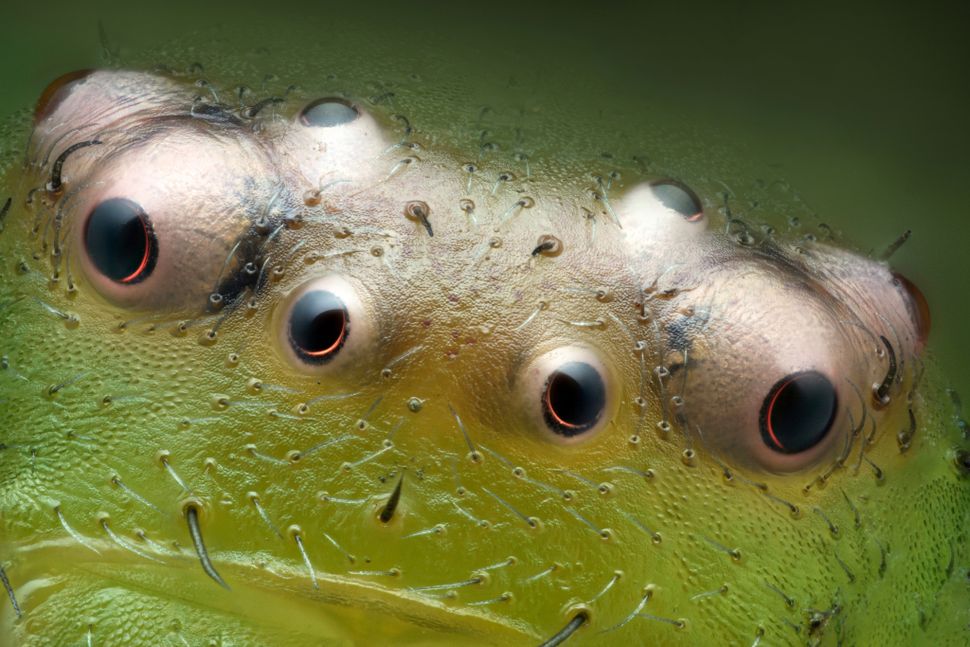
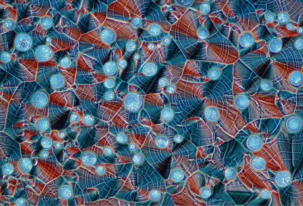
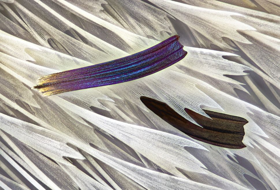
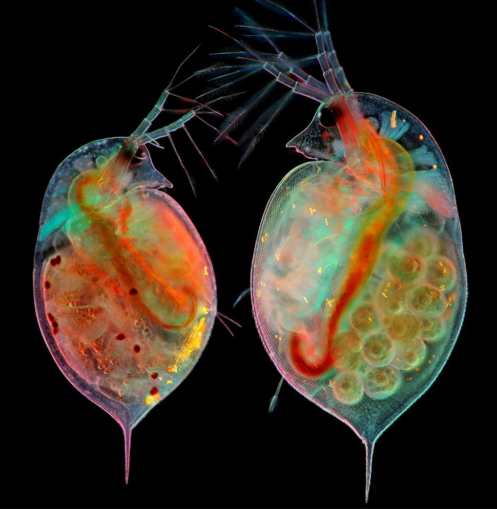
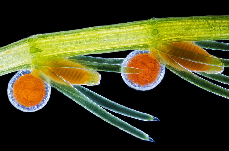
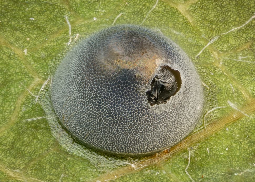
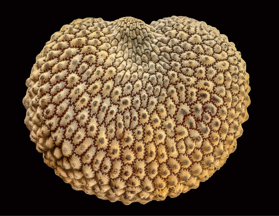
The image of differentiated mouse brain tumor cells taken by Bruno Cisterna and Eric Vitriaol won 1st place in the competition. (Image credit: Dr. Bruno Cisterna & Dr. Eric Vitriol/Nikon Small World 2024)This image by Marcel Clemens shows an electrical arc between a pin and a wire. (Image credit: Dr. Marcel Clemens/Nikon Small World 2024)This image, taken by Chris Romaine, shows the leaf of a cannabis plant. The bulbous glands are trichomes — a small hair or growth from the epidermis of a plant — while the purple bubbles contain cannabinoids. (Image credit: Chris Romaine/Nikon Small World 2024)This image, taken by Amy Engevik, shows a section of a small intestine of a mouse. (Image credit: Dr. Amy Engevik/Nikon Small World 2024)A close-up of a cluster of octopus eggs taken by Thomas Barlow and Connor Gibbons. (Image credit: Thomas Barlow & Connor Gibbons/Nikon Small World 2024)Henri Koskinen’s photograph of the slime mold Cribraria cancellata. (Image credit: Henri Koskinen/Nikon Small World 2024)A photograph showing the cross section of a leaf from the European beach grass Ammophila arenaria taken by Gerhard Vlcek. (Image credit: Gerhard Vlcek/Nikon Small World 2024)Stephanie Huang’s image shows a neuron from the brain of an adult rat. (Image credit: Stephanie Huang/Nikon Small World 2024)John-Oliver Dum’s photograph shows pollen caught in the web of a garden spider. (Image credit: John-Oliver Dum/Nikon Small World 2024)The spores of the black truffle Tuber melanosporum taken by Jan Martinek. (Image credit: Jan Martinek/Nikon Small World 2024)Ferenc Halmos’ image showing water droplets on slime mold on a rotten twig. (Image credit: Dr. Ferenc Halmos/Nikon Small World 2024)This image, taken by Daniel Knop, shows the wing scales of a butterfly on a medical syringe needle. (Image credit: Daniel Knop/Nikon Small World 2024)Paweł Błachowicz captured the eyes of the green crab sider Diaea dorsata. (Image credit: Paweł Błachowicz/Nikon Small World 2024)Marek Miś’ image shows the recrystallized mixture of hydroquinone, a compound that reduces melanin production, and myoinositol, a type of sugar found in the body but also some foods and supplements. (Image credit: Marek Miś/Nikon Small World 2024)Sébastien Malo’s photograph shows the scales of the Madagascan sunset moth wing Chrysiridia ripheus. (Image credit: Sébastien Malo/Nikon Small World 2024)Marek Miś also captured an image of two water fleas with embryos and eggs. (Image credit: Marek Miś/Nikon Small World 2024)Frantisek Bednar’s image shows the reproductive organs of the stonewort algae Chara virgata. (Image credit: Dr. Frantisek Bednar/Nikon Small World 2024)An image of an insect egg parasitized by a wasp taken by Alison Pollack. (Image credit: Alison Pollack/Nikon Small World 2024)Allison Pollack also captured this image of a seed of a silene plant. (Image credit: Alison Pollack/Nikon Small World 2024)Bruno Cisterna and Eric Vitriol also entered this image of an early stage mouse glioblastoma. (Image credit: Dr. Bruno Cisterna & Dr. Eric Vitriol/Nikon Small World 2024)
Scientists convert plastic waste into clean hydrogen using sunlight, offering solutions for pollution and sustainable energy. Solar-powered technology could soon turn plastic waste into clean fuel—tackling pollution and energy in a single bold…
Researchers built a miniature human bladder to demonstrate how the composition of urine weakens bladder tissue, promoting the recurrence of infections even after the use of antibiotics. Animated Z-stack showing the volume of…
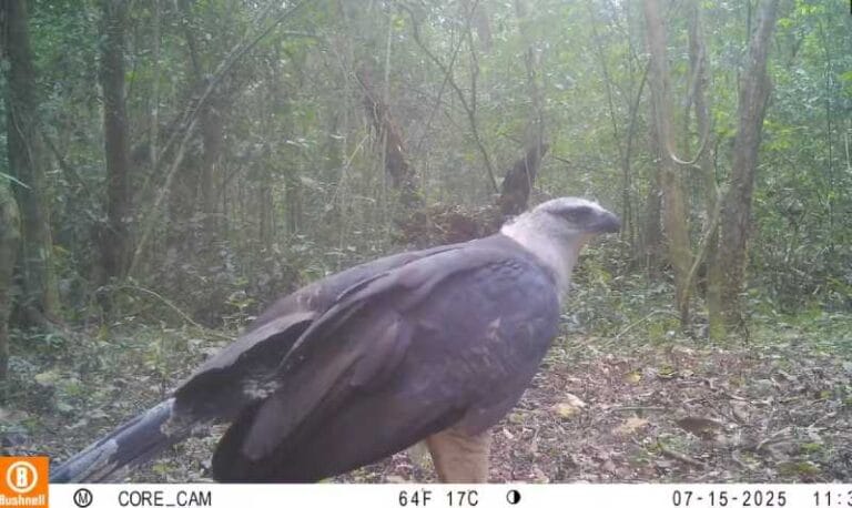
Endangered bird of prey captured by camera trap. The video is unprecedented in the park and may indicate a stable population of the species in the area. Crested eagle (*Morphnus guianensis*) recorded at…
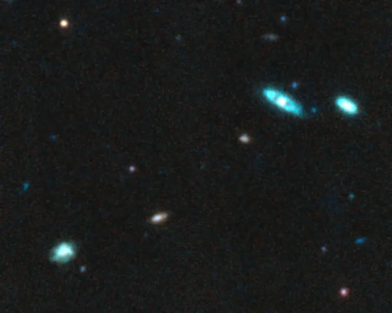
Galaxies at ‘cosmic noon’: Research gives deep dive into universe’s wild growth spurt. Observed MEGA galaxies have a lot of variation in color and morphology, giving insight into the galaxies' ages, dust content…
Инженеры преобразуют клетки кожи напрямую в нейроны для клеточной терапии. Исследователи из MIT разработали упрощенный процесс преобразования клетки кожи непосредственно в нейрон. Это изображение показывает преобразованные нейроны (зелёные), которые интегрировались с нейронами стриатума…